Sclera, Limbus & Clinical Correlations
High-Yield Clinical + PYQ Focus
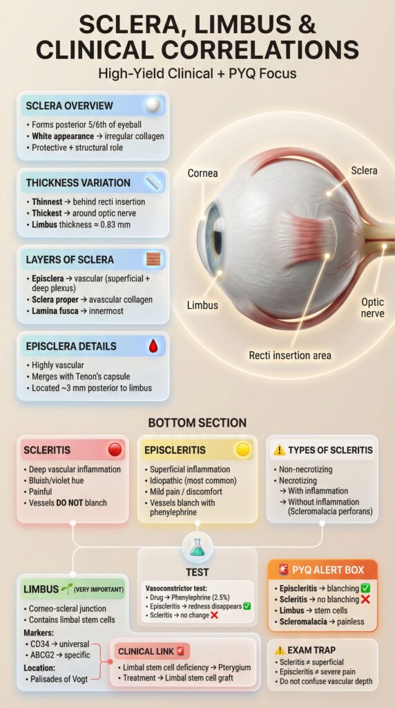
Sclera Overview
• Forms posterior 5/6th of eyeball
• White appearance → irregular collagen fibers
• Protective + structural role
• Non-elastic (unlike cornea)
Thickness Variation
Thinnest: behind recti insertion (0.4 mm)
Thickest: around optic nerve (1 mm)
Limbus: ≈ 0.83 mm
Average: 0.6-0.8 mm
Layers of Sclera
- Episclera → highly vascular (superficial + deep)
- Sclera proper → avascular (main bulk)
- Lamina fusca → innermost pigmented layer
Episclera Details
• Highly vascular (2 plexuses)
• Merges with Tenon’s capsule
• Located ~3 mm posterior to limbus
• Inflammation here → blanches with phenylephrine
Scleritis
• Deep vascular inflammation
• Bluish/violet hue (characteristic)
• Severe pain (can wake at night)
• Vessels DO NOT blanch with drops
• Associated with systemic disease (40-50%)
Episcleritis
• Superficial inflammation
• Idiopathic (most common, 90%)
• Mild pain / Foreign body sensation
• Vessels blanch with phenylephrine
• Self-limited (2-3 weeks typically)
Types of Scleritis
- Non-necrotizing (anterior/posterior)
- Necrotizing with inflammation (painful)
- Scleromalacia perforans (painless, worst prognosis)
Necrotizing types → higher systemic disease risk
Vasoconstrictor Test
Drug: Phenylephrine 2.5%
✔ Episcleritis: redness disappears
✖ Scleritis: redness persists
Gold standard clinical test
Scleritis
Deep inflammation • Painful • No blanchingEpiscleritis
Superficial • Mild pain • Blanches with dropsKey Difference
Depth of inflammation • Vascular response🔬 Limbus (Very Important)
Corneo-scleral junction where magic happens!
📍 Contains: Limbal stem cells (regenerate corneal epithelium)
🧬 Markers:
Location: Palisades of Vogt (radial ridges)
✨ Function: Prevents keratinization, maintains corneal clarity
⚠️ PYQ Alert Box
✔ Episcleritis → redness blanches with phenylephrine
✖ Scleritis → redness DOES NOT blanch
✔ Limbus → contains regenerative stem cells
✖ Scleromalacia → painless but destructive (necrotizing)
🎯 Rule: Scleritis pain severity > Episcleritis always
🎯 Exam Trap & Link
Common Mistakes:
• Scleritis ≠ superficial
• Episcleritis ≠ severe pain
Clinical: Limbal stem cell deficiency → Pterygium | Treatment: Limbal graft